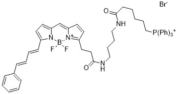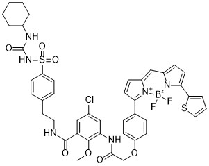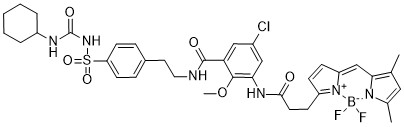



Hoechst 33342, Blue Fluorescent DNA Stain is a cell-permeant fluorescent dye designed for nuclear staining, DNA visualization, and fluorescence microscopy. It selectively binds to the minor groove of double-stranded DNA, particularly AT-rich regions, producing strong blue fluorescence upon DNA binding. Additionally, its membrane-permeable properties enable nuclear staining in living and fixed cells, supporting cell imaging, nuclear morphology analysis, and fluorescence-based biological research.
Key Features and Benefits
- Blue Fluorescent DNA Stain: Exhibits typical excitation near 350 nm and emission near 461 nm when bound to DNA, supporting fluorescence-based nuclear detection.
- DNA Minor Groove Binding: Selectively associates with AT-rich regions of double-stranded DNA, providing strong fluorescence enhancement upon binding.
- Cell-Permeant Fluorescent Dye: Penetrates intact cell membranes, enabling nuclear staining in living cells under suitable experimental conditions.
- High Nuclear Staining Contrast: Produces bright nuclear fluorescence while maintaining relatively low background fluorescence under optimized staining conditions.
- Live-Cell Imaging Compatibility: Specifically supports visualization of cell nuclei in living cells without requiring membrane permeabilization.
- Fixed-Cell Staining Capability: Enables fluorescent nuclear labeling in fixed-cell preparations for microscopy and cellular analysis.
- DNA Content Analysis: Supports fluorescence-based assessment of cellular DNA content and cell cycle distribution using validated staining protocols.
- Fluorescence Instrument Compatibility: Works with fluorescence microscopes, flow cytometers, and imaging systems equipped with appropriate UV excitation and blue emission detection settings.
- Nuclear Research Versatility: Additionally, supports studies of nuclear morphology, cell proliferation, apoptosis-associated chromatin changes, and cellular organization.
Applications
- Fluorescent Nuclear Staining: Enables visualization of cell nuclei in living and fixed cells for fluorescence microscopy and biological imaging.
- Live-Cell Imaging: Supports monitoring of nuclear structures and cellular organization in viable cell populations.
- DNA Visualization: Provides fluorescent labeling of double-stranded DNA for examining nuclear distribution and chromatin organization.
- Cell Cycle Analysis: Specifically supports DNA content measurements for investigating cell cycle distribution using compatible fluorescence detection methods.
- Nuclear Morphology Studies: Enables examination of nuclear size, shape, chromatin condensation, and structural changes.
- Apoptosis Research: Supports identification of apoptosis-associated nuclear condensation and fragmentation in combination with appropriate confirmatory assays.
- Fluorescence Microscopy: Provides bright blue nuclear fluorescence for cellular imaging and multicolor fluorescence experiments.
- Flow Cytometry Applications: Additionally, supports fluorescence-based DNA staining and cellular analysis using suitable excitation and detection settings.
- Cell Proliferation Research: Enables investigation of nuclear characteristics and DNA content changes associated with cellular growth and proliferation.
Storage and Handling
- Storage: Store at −20°C in a tightly sealed container, protected from light and moisture, unless product-specific instructions indicate otherwise. Additionally, avoid repeated temperature fluctuations to preserve dye stability.
- Handling: Use appropriate protective equipment and clean laboratory instruments. Prepare stock solutions in water or DMSO, depending on the supplied formulation and validated solubility.
- Cell Staining: Dilute the dye in a compatible staining buffer and incubate with living or fixed cells. Subsequently, optimize concentration, incubation time, and washing conditions to achieve clear nuclear fluorescence.
- Fluorescence Analysis: Detect DNA-bound fluorescence using suitable UV excitation and blue emission settings. Minimize light exposure and evaluate nuclear staining, background fluorescence, and cell viability using appropriate controls.
Hoechst 33342 Blue Fluorescent DNA Stain combines selective DNA binding with strong blue fluorescence for sensitive nuclear visualization. Therefore, it supports live-cell imaging, DNA content analysis, nuclear morphology studies, cell cycle research, and fluorescence-based cellular investigations.
AxisPharm offers 5000+ PEG Linkers with high purity. Different kinds of PEG Reagents may be available by custom synthesis.