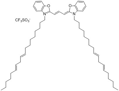



DiI Lipophilic Fluorescent Tracer is a lipophilic carbocyanine fluorescent dye designed for cell membrane labeling, neuronal tracing, and fluorescence imaging. Its hydrophobic alkyl chains promote incorporation into lipid bilayers, while its orange-red fluorescence enables visualization of labeled membranes. Additionally, DiI supports cell tracking, membrane dynamics studies, and cellular morphology research in compatible biological systems.
Key Features and Benefits
- Lipophilic Carbocyanine Dye: Incorporates into lipid membranes through hydrophobic interactions, providing effective membrane-associated fluorescence labeling.
- Orange-Red Fluorescence: Exhibits typical excitation near 549 nm and emission near 565 nm, although spectral properties vary with the environment.
- Membrane Labeling Capability: Specifically enables visualization of cellular membranes, membrane structures, and lipid-associated compartments.
- High Fluorescence Intensity: Provides detectable fluorescence in lipid environments for microscopy and cell imaging applications.
- Neuronal Tracing Compatibility: Supports visualization of neuronal membranes and pathways through membrane-associated dye diffusion.
- Cell Tracking Versatility: Enables fluorescent labeling of cells for studying distribution, migration, and cellular interactions.
- Membrane Retention: Remains associated with lipid-rich structures, although retention and dye transfer depend on experimental conditions.
- Fluorescence Microscopy Compatibility: Supports analysis using fluorescence microscopes equipped with appropriate excitation and emission filters.
- Research Flexibility: Additionally, accommodates applications involving live cells, fixed tissues, and compatible membrane preparations.
Applications
- Cell Membrane Labeling: Enables fluorescent visualization of cell membranes in microscopy and cellular research.
- Neuronal Tracing: Supports investigation of neuronal morphology, membrane projections, and anatomical connectivity.
- Cell Tracking Studies: Facilitates monitoring of labeled cell populations, migration, and distribution in compatible experimental models.
- Fluorescence Microscopy: Specifically supports imaging of membrane structures using appropriate fluorescence detection systems.
- Membrane Dynamics Research: Enables investigation of membrane organization, movement, and interactions in biological systems.
- Cell Morphology Analysis: Supports visualization of cell boundaries, processes, and structural features.
- Tissue Labeling: Enables membrane-associated fluorescent staining of compatible biological tissue preparations.
- Cell Interaction Studies: Additionally, supports investigation of cell distribution and interactions using fluorescent membrane labeling.
- Biomedical Imaging Research: Provides a fluorescent labeling reagent for developing and evaluating experimental cellular imaging methods.
Storage and Handling
- Storage: Store at −20°C in a dry, tightly sealed container, protected from light and moisture. Minimize temperature fluctuations to preserve dye stability.
- Handling: Use appropriate protective equipment and clean laboratory instruments. Prepare stock solutions in compatible solvents, such as DMSO, DMF, or ethanol, after confirming solubility.
- Cell Labeling: Dilute the dye into a compatible labeling medium and optimize concentration, incubation time, and washing conditions. Additionally, minimize light exposure during sample preparation.
- Fluorescence Analysis: Use appropriate orange-red fluorescence detection settings. Subsequently, assess staining uniformity, cell viability, and background fluorescence while accounting for potential dye transfer.
DiI Lipophilic Fluorescent Tracer combines strong membrane affinity with orange-red fluorescence for cellular imaging applications. Therefore, it supports cell membrane labeling, neuronal tracing, cell tracking, and fluorescence-based investigations of cellular organization and behavior.
AxisPharm offers 5000+ PEG Linkers with high purity. Different kinds of PEG Reagents may be available by custom synthesis.